- General ophthalmological examination
- Slit-lamp examination
- Tear film measurement
- Intraocular pressure measurement
- Measurement of pupil size in the dark
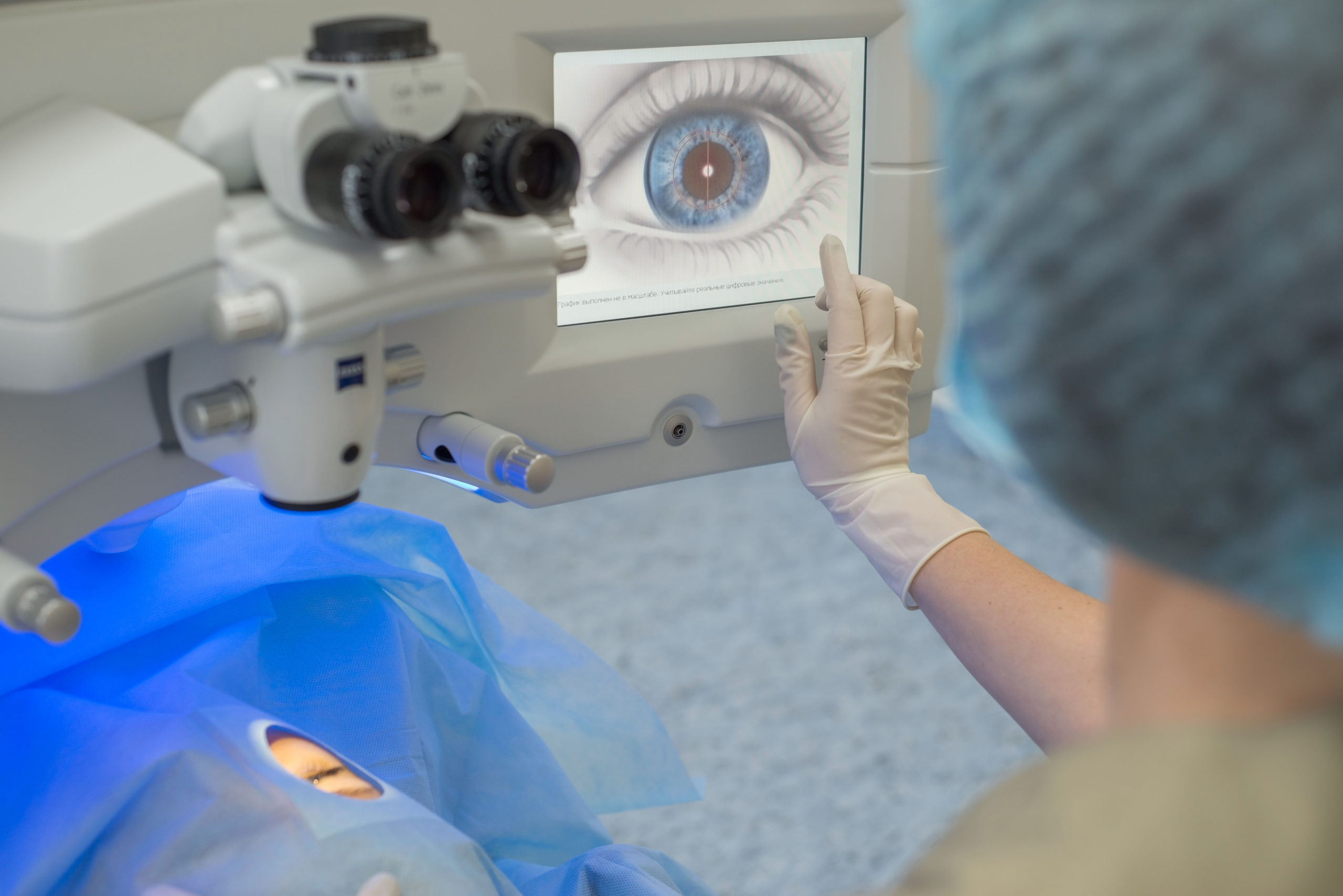
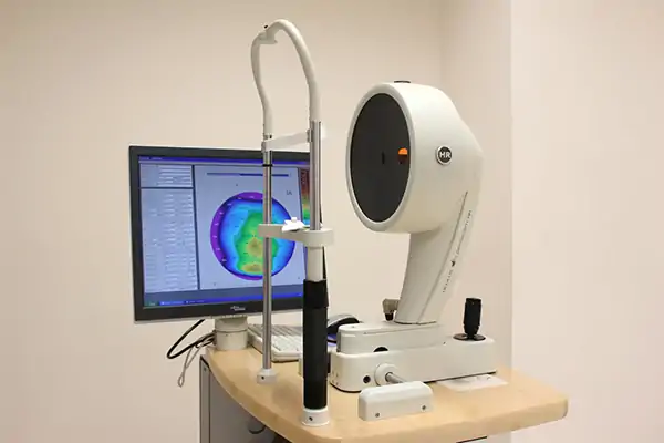
- Corneal topography
At Augenland, we are dedicated to helping people with visual impairments achieve optimal vision through a minimally invasive procedure. That’s why everything at Augenland revolves around providing each patient with personalized care, the highest quality standards, and top medical expertise.
The preliminary examination for eye laser treatment is an essential part of the procedure. It allows the surgeon to determine the exact characteristics of the eye. A thorough preliminary check guarantees safety and precision during the laser treatment.
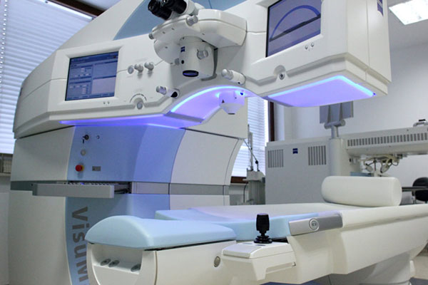
Augenland is equipped with the most advanced diagnostic devices, and here we would like to introduce the most important ones to you.
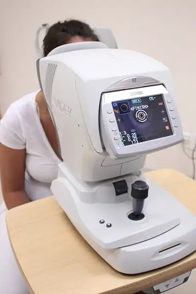
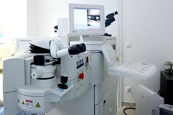
The phoropter enables highly accurate automatic refraction. For seamless data transfer, the phoropter is directly connected to various devices. Under the most natural visual conditions possible, an exact prescription for corrective lenses is determined. For testing near visual acuity, an illuminated near reading chart is integrated. The improved viewing lenses with an anti-smudge coating provide even more precise results.
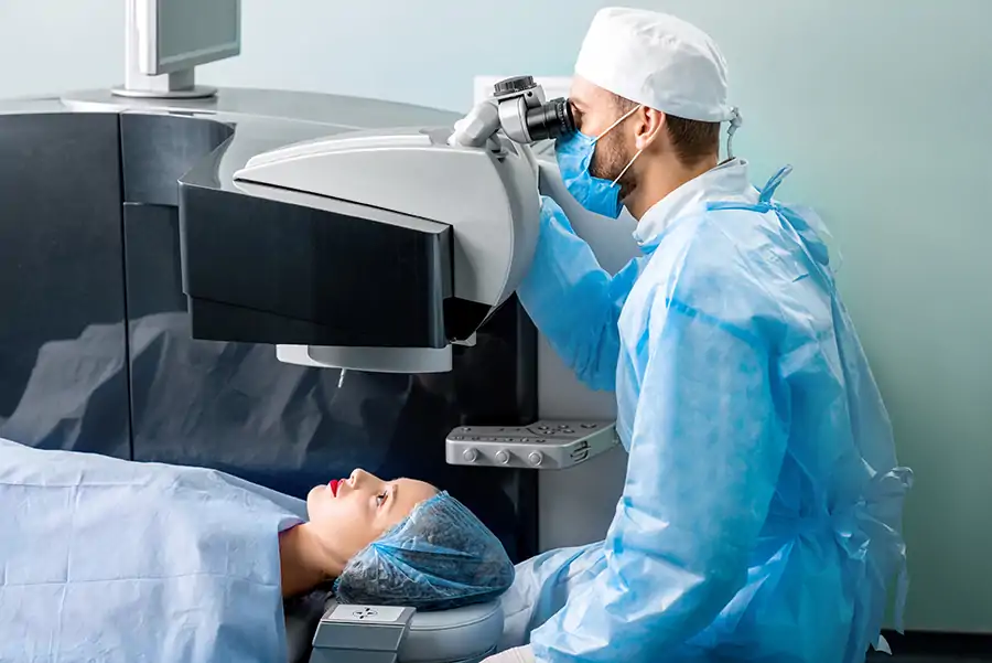
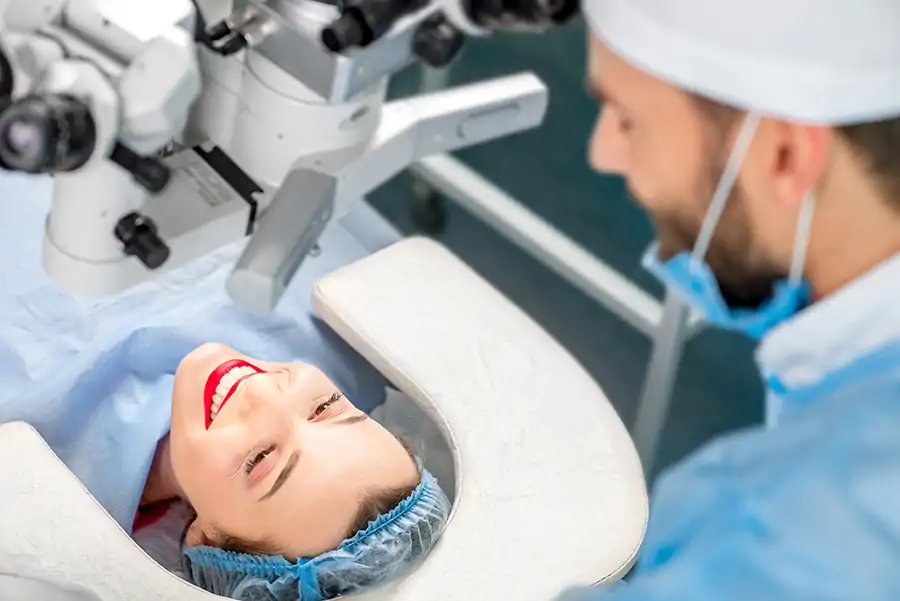
The LASIK procedure is performed without general anesthesia, injections, or other strong anesthetics. A mild sedative tablet and special numbing eye drops are entirely sufficient. Even during the laser treatment, you can communicate with the surgeon. Should your eyes move during the LASIK procedure, the eye tracker will detect it. The eye tracker follows even the tiniest eye movements, ensuring that the laser treatment is always carried out precisely in the correct spot.
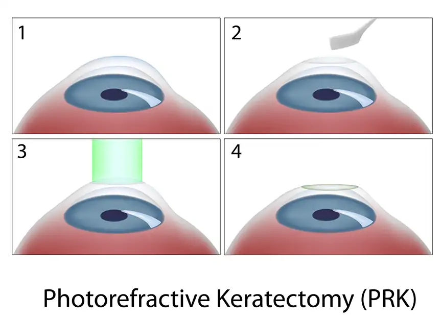
The cornea is located at the front of the eye, is transparent, and shaped like a watch glass. It consists of non-vascular tissue, is usually about half a millimeter thick, and functions as a powerful lens. Once the cornea has been gently fixed in place with a silicone ring—completely without pain—the femtosecond laser separates a very thin, circular corneal flap, about one-tenth of a millimeter thick. This flap is then lifted by the surgeon.
The now-exposed corneal layer is optimized with the excimer laser through a computer-controlled, targeted removal of tissue. By altering the cornea’s refractive power, the refractive error is corrected. The actual laser process takes only a few seconds and is completely painless. Any unintended eye movements are immediately detected by the eye tracker system and compensated for by the laser. Finally, the corneal flap is folded back into place. Due to adhesion forces, it immediately rests securely and precisely in its original position.
Every patient is treated and examined using the best available software and hardware. All advanced functions such as WaveScan, iris registration, eye tracker, and tissue-saving technology are utilized.