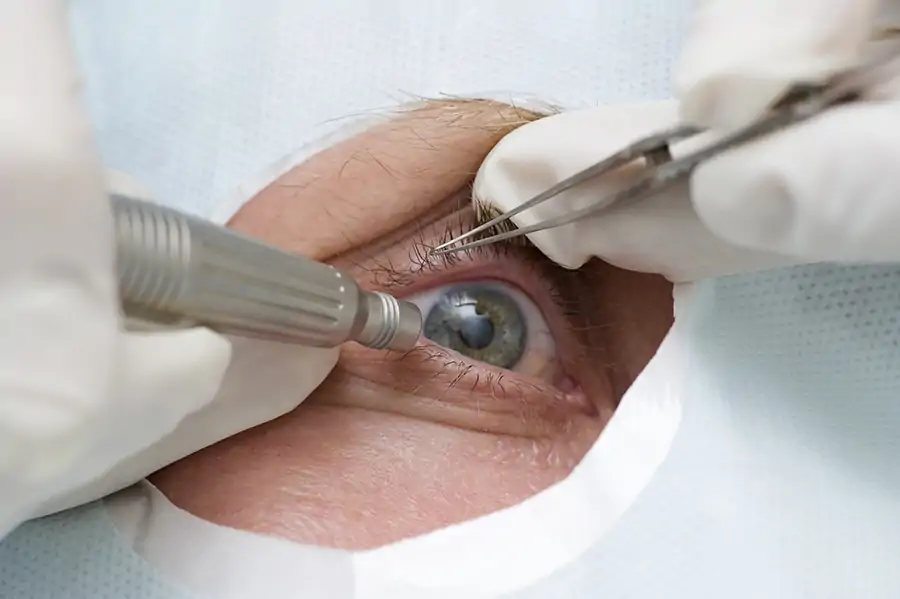
The ultrasound examination is a frequently used procedure that is also used in ophthalmology. Ultrasound devices are not dangerous for the patient and side effects are excluded. The probe head, a pen-like probe, emits sound waves; these hit a surface and are reflected and received again by the probe. This creates an image of the examined tissue.
In ophthalmology there are two types of ultrasound devices. With the A-image of the ultrasound, the ophthalmologist measures the axial length of the eye and the radius of the cornea. These measurements are important in a cataract operation; with them the artificial lens to be inserted can be calculated.
With the B-image, pathological changes or tumors can be detected.
Simply contact us via our website, by telephone or via WhatsApp – we look forward to seeing you!

