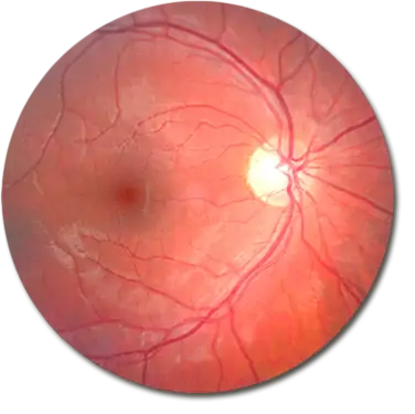
To identify the type of macular degeneration, Augenland uses what is known as fluorescence angiography. This examination allows the fine blood vessels of the retina to be displayed in excellent detail. For this purpose, a dye (fluorescein) is injected into a vein in the arm. The dye is distributed through the bloodstream throughout the body, including the blood vessels of the eye. A special camera then captures the blood vessels of the eyes.
This examination is an important diagnostic tool for later selecting an appropriate therapy.
For this examination as well, the ophthalmologist must dilate the pupils with special eye drops. Usually, both eyes are examined, even if only one eye shows typical visual disturbances.
First, the ophthalmologist examines the back of the eye. Then, the dye is injected into the arm vein. From there, it reaches the blood vessels of the eyes in about 10–30 seconds. Using a special fundus camera, the doctor takes several consecutive images. After about 10 minutes, a few more images are taken. This series of images shows how the blood flows through the vessels of the retina and whether any blood or fluid is leaking.
Make an appointment today! We will be happy to advise you in a personal consultation!